>> Time Lapse Microscopy << |
||
The communication of cells with patterned surfaces is a dynamic process. Therefore it is neccessary to investigate this communication over some time. Even if the parameter derivation from adhesion patterns or cell protein distribution works well on fixed specimen the living state is of higher information content. Time Lapse Microscopy (TLM) requires increased attention. Besides the higher level of instrumentation power required especially the long investigation times for every experiment must be considered. Further, the amount of information from every single experiment is enormous. Time lapse microscopy needs the experimentators engagement in both instrumentation and data evaluation. |
||
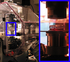
Far the most cells need a carefull environment control. A temperature too low stops cell activity. Too high temperatures (even over short times) can stress or kill cells. The humidity must be high to prevent up concentration of ions by medium dessication. A carbon dioxide supply stabilizes the medium pH. A constant temperature also prevents optical defocussing by thermic activity of the microscope. Ease of handling is not the most important topic in time lapse microscopy. As the effort also the results of time lapse microscopy are outstanding. Lets have a look to the second figure. This shows a CHO epithelial cell after vital staining with Calcein and TIRF microscopy. The furrows are characteristic. Now lets click on the picture to open the animation. The furrows are no static structures. Even cells which do not migrate (like CHO) permanently reorganize the furrow pattern. In the cell periphery the turn over is increased as compared to the cell center. Also the actin cytosceleton is under permanent reconstruction as can be seen in the third figure. Here a C2C12 muscle cell is gentechnologically enabled to produce fluorescent actin protein. Consequently upon excitation the cells cytosceleton dynamic can followed over time. after clicking on the figure the animantion shows that the fibres are prolonged at the ends. During migration the C2C12 cell extends along the fibers. Time lapse microscopy can show how migrating cells react to surface patterns (last figure). Again clicking on the figure opens an animation. The pattern is so attractive that intercellular connections are disrupted As a result one cell remains on a structure pad while another one migrates to the right. Time lapse microscopy makes it possible to look to the communication process between adherent cells and patterned surfaces. The results are worth to invest time in the experiments and the data evaluations. Otherwise results always have the character of snapshots. |
||||
 |
||||
 |
||||
 |
||||
 |
||||
 |
 |
 |
||